Blog
CT Scan vs. MRI Scan in Dogs and Cats: What’s the Difference?
Computed tomography (CT) and magnetic resonance imaging (MRI) are among the most advanced diagnostic imaging methods in modern veterinary medicine. They help veterinarians obtain timely and accurate diagnoses for many conditions in companion animals. Both imaging techniques provide detailed views of the body’s internal structures, but each has distinct advantages and is used for different clinical indications.
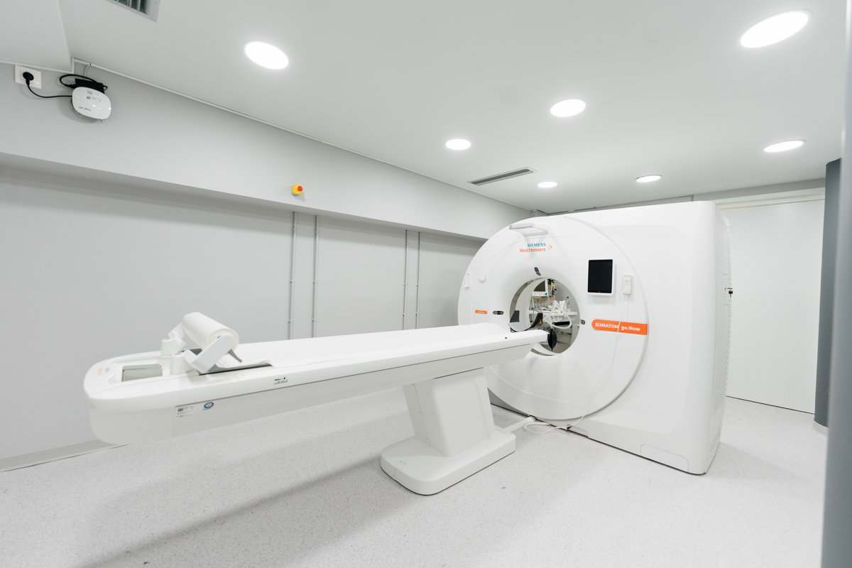
Computed Tomography (CT)
Computed tomography (CT) is an advanced imaging technique that uses X-rays to produce detailed images of the body’s internal structures. Specialized software combines these images to create three-dimensional representations of organs, bones, and tissues.
In veterinary medicine, CT scans are frequently used to evaluate injuries, fractures, and bone disorders. They are also particularly useful for assessing conditions affecting the nasal cavity, chest, and lungs, as well as for detecting tumors in the abdominal or thoracic regions.
One of the main advantages of CT scanning is that the examination is completed very quickly. This reduces the duration of anesthesia, which is particularly important for the animal’s safety.

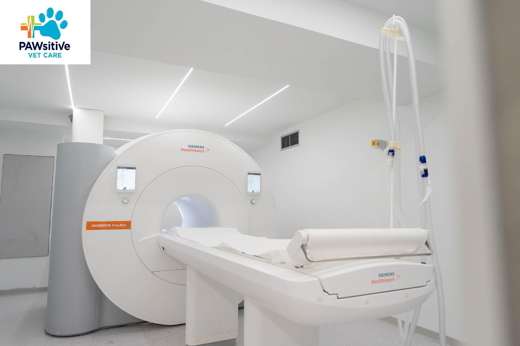
Magnetic Resonance Imaging (MRI)
Magnetic resonance imaging (MRI) uses a strong magnetic field and radio waves to produce highly detailed images of the body’s soft tissues. Unlike CT scans, MRI does not use ionizing radiation.
MRI is considered the most reliable imaging method for evaluating the brain, spinal cord, and the nervous system in general. For this reason, it is often used to investigate neurological problems in dogs and cats, such as seizures or paralysis.
In addition, MRI provides exceptionally detailed images of muscles, ligaments, and other soft tissues, helping veterinarians diagnose injuries that may not be easily detected with other imaging methods.
Key Differences Between CT and MRI
CT scans rely on X-rays and are particularly effective for imaging bones and lungs. The examination is completed very quickly, which is why CT is often used in emergency situations or to assess traumatic injuries.
Magnetic resonance imaging (MRI), on the other hand, uses a magnetic field and provides superior visualization of soft tissues, including the brain and spinal cord. The examination takes longer but offers greater detail.

Anesthesia Requirements
In most cases, both CT and MRI scans are performed under general anesthesia. This is necessary because the animal must remain completely still during the examination in order to obtain clear and accurate images.
Anesthesia is administered under the supervision of a specialized veterinarian from our team, with continuous monitoring of the animal’s vital signs.